Overview
An X-Ray is a quick, painless test that produces images of the structures inside your body, particularly your bones. The X-Ray machine produces safe level of radiation through your body, and they are absorbed in different amounts depending on the density of the material they pass through. Dense materials, such as bone and metal, show up as white on X-Rays. The air in your lungs shows up as black. Fat and muscle appear as shades of grey. For some types of X-Ray tests, a contrast medium such as iodine or barium is introduced into your body to provide greater detail on the images. Some people worry that X-Rays aren’t safe because radiation exposure can cause cell mutations that may lead to cancer. The amount of radiation you’re exposed to during an X-Ray depends on the tissue or organ being examined. However, radiation exposure from an X-Ray is negligible, and the benefits from these tests far outweigh the risks.
Why an X-Ray is done?
- Fractures and infections – In most cases, fractures and infections in bones and teeth show up clearly on X-rays.
- Arthritis – X-rays of your joints can reveal evidence of arthritis. X-rays taken over the years can help your doctor determine if your arthritis is worsening.
- Dental decay – Dentists use X-rays to for cavities in your teeth.
- Osteoporosis – Special types of X-ray tests can measure your bone density.
- Bone cancer – X-rays can reveal bone tumors.
- Lung infections or conditions – Evidence of pneumonia, tuberculosis or lung cancer can show up on chest X-rays.
- Breast cancer – Mammography is a special type of X-ray test used to examine breast tissue.
- Enlarged heart – This sign of congestive heart failure shows up clearly on X-rays.
- Blocked blood vessels – Injecting a contrast material that contains iodine can help highlight sections of your circulatory system to make them visible on X-rays.
- Digestive tract problems – Barium, a contrast medium delivered in a drink or an enema, can help reveal problems in your digestive system.
- Swallowed items – If your child has swallowed something such as a key or a coin, an X-ray can show the location of that object.
How to Prepare for an X-ray
Preparation for an X-ray is generally minimal, but it can vary depending on the area of the body being examined.
- Remove Metal Objects: You will be asked to remove any jewelry, eyeglasses, removable dental appliances, and other metal objects from the area being X-rayed, as these can block the X-ray beam and obscure the image.
- Wear Loose Clothing: You may be asked to change into a hospital gown, especially if the clothing you are wearing contains metal components like zippers, snaps, or buttons.
- Inform Your Doctor: It is crucial to tell your doctor or the technologist if you are or might be pregnant. X-rays are a form of radiation, and while the dose is small, special precautions may be taken to protect a developing fetus.
- Specific Instructions: For certain types of X-rays, such as those of the digestive tract, you may be asked to fast or follow a specific diet beforehand. If a contrast medium (a special dye) is used to highlight a specific area, you may need to drink a solution, get an injection, or receive an enema.
How it’s Done
The CT scan procedure is a quick and straightforward process managed by a trained technologist.
 1
1
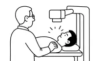
Positioning:
You are asked to lie still on the examination table.
 2
2
Staying Still:
You must remain still for some scans, you may be asked to hold your breath briefly.
 3
3
Protection:
A lead apron or shield is placed to protect areas not being imaged from radiation.
 4
4
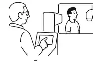
The Exposure:
The technologist will reviews the images on a computer or may take several images from different angles to get a complete picture.
How it will Feel
The procedure itself is painless and you will not feel the X-ray radiation passing through your body. The only sensations you might feel are related to the positioning.
- Discomfort from Positioning – Depending on the body part being imaged and your physical condition (e.g., if you have an injury or arthritis), you may experience brief discomfort from holding a specific position on the hard table or plate. The technologist will do their best to make you as comfortable as possible.